How to Prevent Blood Clots When Traveling Long Distances Introduction...
Read More


How Smoking Damages Blood Vessels and What Happens When You Quit
How Smoking Damages Blood Vessels and What Happens When You...
Read More
Personalized Anticoagulation Strategies in Patients with Venous Thromboembolism
Personalized Anticoagulation Strategies in Patients with Venous Thromboembolism: Outline Venous...
Read More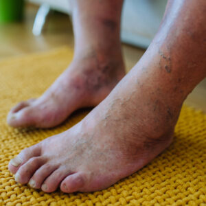
Managing Complex Peripheral Artery Disease in Diabetic Patients
Managing Complex Peripheral Artery Disease in Diabetic Patients Introduction Peripheral...
Read More
The Link Between Smoking and Vascular Disease
The Link Between Smoking and Vascular Disease Smoking is one...
Read More
How Vascular Health Changes with Age
How Vascular Health Changes with Age Aging is a natural...
Read More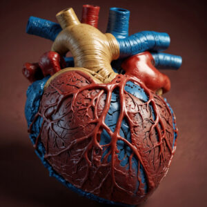
Understanding the Circulatory System
Understanding the Circulatory System: How It Affects Your Health The...
Read More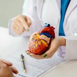
What Are Vascular Diseases
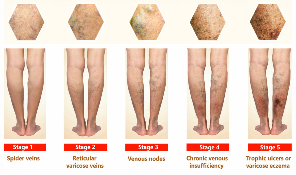
What Are Vascular Diseases? Symptoms, Causes, and Risk Factors Introduction...
Read More